
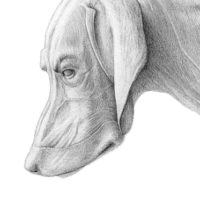
Species-specific accuracy
Illustrations developed from anatomical references, scientific source material, and species-specific structures.
Clarity for complex animal anatomy
Visuals designed to explain layered anatomy, organ systems, musculoskeletal structures, and functional relationships.
Structured visual communication
A clear workflow from reference material and research to final visuals for education, publication, or veterinary use.
Animal Anatomy Illustration Services
Animal anatomy illustration translates complex anatomical information into clear, accurate visual communication for research, education, veterinary medicine, and scientific publishing. Whether the subject is a mammal, a bird, a fish, a reptile, or a comparative anatomical structure, the goal is always the same: to make biological complexity understandable without sacrificing scientific accuracy.
Photography, scans, and reference material can provide valuable information, but they rarely communicate structure with complete clarity. Animal anatomy often involves layered tissues, subtle proportions, species-specific variation, and internal relationships that are difficult to explain through raw images alone. Illustration clarifies, organizes, and presents these structures with purpose.
I am Karin Spijker, a certified scientific and medical illustrator with a strong background in anatomy, drawing, and biological visualization. I create custom animal anatomy illustrations for educational materials, scientific publications, veterinary communication, and research-based visual projects.
Related pages:
→ anatomy illustration
→ veterinary medical illustration
→ scientific illustration
→ portfolio
“I have worked with Karin Spijker for many years on various visual projects. The greatest common denominator in these projects is a qualitative, professional image delivered on time. Karin is entirely at home in both fields, whether an illustration or high-end image editing on photo material.”
“Karin Spijker has performed assignments for me several times to my complete satisfaction, such as logos and two 3D animations. Karin can translate the information from a briefing into the desired end product and can think along with you pleasantly. In doing so, she works accurately, follows the set timetable, and honors her appointments. Karin is also a charming person to work with.”
 Natasja Kardos, Seahorse Solutions
“For my clients, I have asked Karin Spijker more often for customized assignments, especially for more specialized image editing. Karin can conjure up software like Adobe Photoshop and Illustrator. She is very meticulous and also communicates about progress. I highly recommend Karin, her work, and her pleasant cooperation.”
 Marian de Jong, Piri Piri Marketing en Communicatie
Translating animal anatomy into visual clarity
An animal anatomy illustration is not simply about drawing animals. It is about translating anatomical knowledge into reliable visual information. In veterinary education, zoology, comparative anatomy, and life science communication, visual clarity directly influences how information is understood and applied.
A well-developed animal anatomy illustration can isolate important structures, remove unnecessary visual noise, and guide the viewer toward the relationships that matter most. This is especially valuable when explaining internal anatomy, musculoskeletal systems, organ structures, vascular relationships, or species-specific anatomical differences.
Unlike generic animal artwork, scientific animal anatomy illustration is built around accuracy, structure, and communication. The visual style may range from clean line work to detailed digital rendering or 3D visualization, but the foundation remains the same: the anatomy must be correct, readable, and aligned with the project’s purpose.
What animal anatomy illustration is used for
- veterinary education and training
- zoological and biological research
- comparative anatomy studies
- scientific publications and textbooks
- educational platforms and museum communication
- veterinary patient or client communication
Scientific accuracy across species
Animal anatomy requires careful attention to species-specific structure. A dog, horse, cat, bird, or fish cannot be visualized as a generic animal body with minor adjustments. Each species has its own proportions, skeletal organization, muscular structure, organ placement, and functional anatomy.
This is where custom illustration becomes important. A professional animal anatomy illustration can be developed from anatomical references, scientific literature, veterinary source material, specimen studies, imaging, or expert input. The final image is not a decorative interpretation but a structured visual explanation grounded in the subject’s biological reality.
This level of accuracy is especially important in comparative anatomy, where differences between species are central to the message. It is also essential in veterinary and educational contexts, where unclear or inaccurate visuals can lead to misunderstanding.
Workflow: from reference material to final illustration
Creating a high-quality animal anatomy illustration requires a structured and transparent process. Each project begins with the communication goal: what needs to be understood, who the audience is, and where the final visual will be used.
From there, relevant references are reviewed. These may include anatomical literature, veterinary sources, photographs, sketches, imaging, scientific papers, or expert input. The concept phase defines the visual hierarchy, composition, and level of detail before the final illustration is developed.
- briefing and scope definition
- research and reference review
- concept sketches and visual planning
- illustration development and refinement
- final delivery for publication, presentation, or digital use
A transparent workflow designed for accuracy, clarity, and efficient collaboration.

From complex biological information to clear visuals
Many animal anatomy projects begin with a specific communication challenge. A researcher may need to explain a structural difference between species. A publisher may need a clear anatomical figure for a textbook. A veterinary educator may need a visual that explains anatomy in a way students can understand quickly.
Illustration makes this possible by applying visual hierarchy. Instead of showing everything at once, it focuses attention on the structures and relationships most relevant to the communication goal. This may involve simplifying background details, separating overlapping structures, clarifying depth, or combining multiple references into a single coherent visual.
A strong animal anatomy illustration can show an “idealized” version of a structure while still remaining scientifically accurate. This is particularly valuable when photography is too noisy, too specific, or unable to clearly show internal organization.
Custom animal anatomy illustration versus stock and AI-generated visuals
- species-specific anatomical accuracy
- full control over structure, detail, and emphasis
- consistency across larger publications or learning materials
- clearer licensing and usage arrangements
- the ability to revise and refine based on expert feedback
About Karin Spijker
I am Karin Spijker, a scientific and medical illustrator with a background in drawing, painting, and textile design. With a master’s in scientific illustration and additional skills in 3D production, I combine accuracy, artistry, and storytelling. My work helps healthcare and publishing teams communicate complex ideas clearly and reliably.
Alongside commissioned projects, I also create independent artworks inspired by nature, anatomy, and landscapes. My mission is to make science and nature accessible, inspiring, and visually engaging.

Why work with a scientific animal anatomy illustrator
An animal anatomy illustration requires more than drawing ability. It requires an understanding of anatomy, scientific interpretation, visual discipline, and the ability to translate complex structures into clear communication.
My work combines a background in medical and scientific illustration with traditional drawing skills, digital techniques, and an interest in animal anatomy, nature, and biological form. This allows me to create visuals that are both scientifically grounded and visually clear.
Projects are developed with attention to accuracy, collaboration, and usability. Whether the final illustration is intended for education, research, publishing, veterinary communication, or a broader scientific audience, the aim is to create work that supports understanding and can be used with confidence.
Animal anatomy illustration for education
Education is one of the most important uses of animal anatomy illustration. Students and learners often struggle with structures that are hidden, layered, or difficult to interpret from real specimens or photographs. Illustrations can reduce cognitive load by organizing information into clear, readable visuals.
For veterinary schools, biology programs, online courses, and educational publishers, animal anatomy illustrations can help explain musculoskeletal systems, organ systems, comparative structures, and functional relationships. They can also be adapted for different learning levels, from introductory education to advanced academic material.
This makes animal anatomy illustration a long-term asset. A well-made visual can be reused across lectures, handouts, textbooks, presentations, e-learning modules, and digital platforms.
Related pages:
→ scientific illustration
→ scientific nature drawing
Veterinary and clinical communication
In veterinary contexts, clear anatomical visuals can support both professional training and communication with animal owners. Complex conditions, procedures, injuries, or anatomical explanations may be difficult to understand through words or diagnostic images alone.
Animal anatomy illustrations can help clarify structures, explain surgical or clinical concepts, and support patient education in veterinary care. The tone and level of detail can be adjusted depending on whether the audience is a veterinary specialist, a student, a publisher, or an animal owner.
This is especially useful when the goal is not to overwhelm the viewer but to provide a calm, clear explanation of what is happening anatomically.
Related page:
→ veterinary medical illustration


Specialized animal anatomy focus areas
- mammalian anatomy
- canine anatomy
- feline anatomy
- equine anatomy
- avian anatomy
- fish anatomy
- comparative anatomy
- veterinary anatomy
Understanding animal anatomy illustration pricing
Every project is custom and depends on scope, complexity, and intended usage. If you would like a clear overview of how pricing is structured, including typical ranges and licensing, you can explore the pricing page.
Long-term value of custom animal anatomy illustration
A strong animal anatomy illustration is not a one-time decorative asset. It can become part of a larger educational or scientific communication system. The same visual approach can be adapted across publications, presentations, learning platforms, posters, digital content, and future project materials.
Custom illustration also allows consistency. When multiple visuals are created for the same publication, species group, or educational course, style, labeling, hierarchy, and anatomical interpretation can remain aligned across the entire project.
This makes custom animal anatomy illustration a strategic investment in clarity, reliability, and professional communication.
Frequently asked questions
Find answers to common questions about the illustration process, timelines, pricing, licensing, file delivery, and collaboration.
Start your animal anatomy illustration project
If you are looking for an animal anatomy illustrator for research, education, veterinary communication, or publication, the focus should always be on clarity, accuracy, and usability.
Whether your project involves a single anatomical figure, a series of educational visuals, or a broader scientific communication project, I can help translate complex animal anatomy into clear, reliable, and professionally prepared visuals.
→ View related anatomy work in the portfolio
→ Get in touch to discuss your project
