
Oncology-focused accuracy
Illustrations developed from validated oncology references, imaging, and clinical source material.
Clarity for complex cancer communication
Structured visuals designed to simplify pathways, tumor biology, and treatment processes.
Publication-ready visualization
Medical visuals created for oncology research, education, pharma communication, and clinical use.
Oncology medical illustration
If you work in oncology, whether in research, clinical practice, publishing, or pharma, you already know one thing: clarity is not optional.
Cancer-related processes are among the most complex in medicine. Tumor biology, immunotherapy, molecular pathways, and surgical interventions all operate across multiple levels of abstraction.
This is where oncology medical illustration becomes essential.
A well-designed visual does not just “show” information; it structures it. It removes noise, guides attention, and translates complexity into understanding.
My name is Karin Spijker. As a specialized medical illustrator, I work with researchers, healthcare professionals, and life science companies to translate complex oncological data into clear, scientifically grounded visualizations.
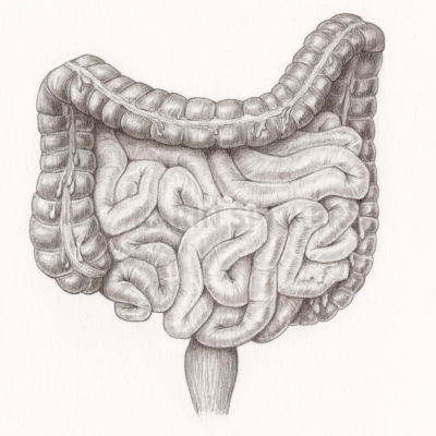
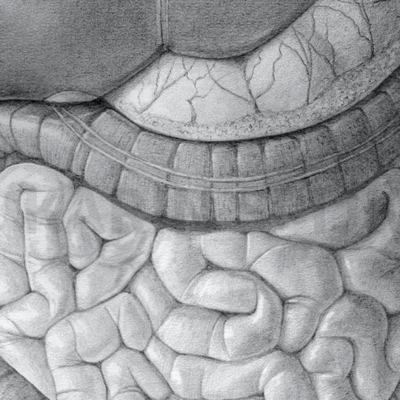
Whether your focus is breast cancer, colon cancer, or intestinal cancer, the goal is always the same:
to create visuals that are accurate, usable, and aligned with the highest professional standards.
Related:
→ surgical illustration
→ patient education illustration
→ medical animation
“I have worked with Karin Spijker for many years on various visual projects. The greatest common denominator in these projects is a qualitative, professional image delivered on time. Karin is entirely at home in both fields, whether an illustration or high-end image editing on photo material.”
“Karin Spijker has performed assignments for me several times to my complete satisfaction, such as logos and two 3D animations. Karin can translate the information from a briefing into the desired end product and can think along with you pleasantly. In doing so, she works accurately, follows the set timetable, and honors her appointments. Karin is also a charming person to work with.”
 Natasja Kardos, Seahorse Solutions
“For my clients, I have asked Karin Spijker more often for customized assignments, especially for more specialized image editing. Karin can conjure up software like Adobe Photoshop and Illustrator. She is very meticulous and also communicates about progress. I highly recommend Karin, her work, and her pleasant cooperation.”
 Marian de Jong, Piri Piri Marketing en Communicatie
What oncology medical illustration actually does
Oncology medical illustration is not simply about depicting organs or tumors. Its primary role is to translate highly complex biological and clinical information into clear visual communication that can be understood more efficiently across research, education, pharma, and clinical environments.
Cancer processes often operate across multiple levels simultaneously. Tumor biology involves interactions between cellular behavior, molecular signaling, immune response, vascularization, metastasis, and treatment effects. Raw medical data such as pathology reports, imaging studies, molecular diagrams, or research figures can quickly become visually overwhelming, even for professional audiences.
Medical illustration introduces structure, hierarchy, and selective emphasis. Rather than presenting all available information at once, it focuses on the relationships and mechanisms most important to the communication goal.
In oncology, illustrations are frequently used to clarify:
- how tumors develop and interact with surrounding tissue
- the role of the tumor microenvironment (TME)
- metastasis through vascular or lymphatic systems
- immunotherapy and targeted treatment pathways
- molecular and cellular mechanisms involved in cancer progression
- surgical approaches and treatment strategies
This level of visual clarification is particularly valuable when communicating highly abstract concepts such as immune response, receptor interactions, signaling pathways, or Mechanisms of Action (MoA) for emerging therapies.
For researchers and pharmaceutical communication, oncology visuals help transform complex scientific information into structured communication that supports understanding, presentations, publication, and interdisciplinary collaboration. In clinical and patient-facing contexts, illustration can also reduce confusion and improve understanding during discussions involving diagnosis, procedures, or treatment options.
A strong oncology medical illustration does not simplify the science itself. It organizes complexity into visuals that are clearer, more accessible, and easier to interpret.
See also:
→ biomedical illustration
→ scientific animation visualization
→ anatomy illustration
Why precision is non-negotiable in oncology
- misunderstanding in clinical education
- incorrect interpretation of research
- reduced credibility in publications
maximum scientific accuracy + controlled visual clarity
- high-impact journal publications
- clinical decision-making
- patient communication under emotional pressure
Where oncology illustration creates real impact
In practice, oncology visuals are used across multiple high-stakes contexts.
In research, they strengthen publications, especially graphical abstracts and complex figures.
In pharma, they explain Mechanisms of Action (MoA) for therapies such as immunotherapy, CAR-T, or targeted treatments.
In clinical environments, they support surgical planning and help explain procedures clearly.
And in patient communication, they reduce uncertainty by making abstract processes tangible.
See also: → medical illustration services
Workflow: structured, efficient, and transparent
- Intake and scope definition
- Research and validation
- Concept and sketch phase
- Feedback and refinement
- Final production
- Delivery in required formats
A transparent workflow designed for accuracy, clarity, and efficient collaboration.

Types of oncology visuals I develop
Oncology visualization covers a wide range of medical and scientific communication needs, depending on the clinical, educational, or research context. Some projects focus on clarifying anatomy, while others involve highly abstract biological mechanisms operating at the cellular or molecular level.
For research and pharmaceutical communication, oncology medical illustrations are often used to visualize tumor pathways, molecular interactions, and Mechanisms of Action (MoA) for targeted therapies, immunotherapy, or precision medicine approaches. These visuals help translate highly specialized scientific information into structured communication that can be understood more efficiently by researchers, clinicians, investors, and healthcare professionals.
In surgical oncology and clinical communication, illustrations may clarify tumor localization, procedural approaches, resection margins, or treatment pathways. This is particularly valuable when communicating complex procedures or multidisciplinary treatment strategies.
Oncology visuals are also widely used in patient communication and education. Cancer diagnoses and treatment processes are often emotionally overwhelming, especially when discussions involve anatomy, metastasis, immunotherapy, or treatment effects that are difficult to visualize through text alone. Structured medical visuals can support understanding while reducing unnecessary confusion and visual overload.
Depending on the project, visuals may include:
- anatomical and histological visualization
- molecular and cellular pathway communication
- immunotherapy and targeted treatment visualization
- surgical oncology procedures
- patient education and explanatory graphics
- publication-ready scientific figures and diagrams
Each project is tailored to the communication goal, audience, and level of scientific complexity involved.
About Karin Spijker
I am Karin Spijker, a scientific and medical illustrator with a background in drawing, painting, and textile design. With a master’s in scientific illustration and additional skills in 3D production, I combine accuracy, artistry, and storytelling. My work helps healthcare and publishing teams communicate complex ideas clearly and reliably.
My work focuses on translating complex oncology and biomedical information into structured visual communication for research, education, and clinical use.
Alongside commissioned projects, I also create independent artworks inspired by nature, anatomy, and landscapes. My mission is to make science and nature accessible, inspiring, and visually engaging.

Why clients choose to work with me
Clients in oncology typically look for more than visual execution.
They need a partner who understands both science and communication.
- medically informed decision-making
- strong visual hierarchy and clarity
- ability to translate complex data into structured visuals
- efficient collaboration with researchers and clinicians
Comparison: Custom illustration vs. stock visuals
When working in oncology, the choice of visual approach directly affects the quality and credibility of your output.
Here is the practical difference:
|
Criteria
|
Custom Oncology Illustration (Karin Spijker)
|
Stock / Template Visuals
|
|---|---|---|
|
Scientific accuracy
|
Built from your data, literature, and expert input
|
Generic, often not fully accurate
|
|
Relevance
|
Tailored to your exact case or study
|
Rarely fits your specific context
|
|
Visual clarity
|
Structured hierarchy and guided focus
|
Often cluttered or visually flat
|
|
Collaboration
|
Direct 1-on-1 with a specialist
|
No content-level support
|
|
Usage rights
|
Clear, flexible licensing for professional use
|
Often restricted or unclear
|


Understanding oncology medical illustration pricing
Every project is custom and depends on scope, complexity, and intended usage. If you would like a clear overview of how pricing is structured, including typical ranges and licensing, you can explore the pricing page.
Frequently asked questions
Find answers to common questions about the illustration process, timelines, pricing, licensing, file delivery, and collaboration.
Conclusion
Oncology demands communication that is as precise as the science itself.
A strong oncology medical illustration bridges the gap between complex data and real understanding. It strengthens publications, supports clinical decisions, and improves communication across all levels.
If you are working on a project involving breast cancer, colon cancer, or intestinal cancer, and need visuals that are both accurate and effective, contact
Get in touch to discuss your project or request a tailored quote.
You can also explore the portfolio to see how complex medical content is translated into clear, reliable visuals.
