
Canine anatomical accuracy
Illustrations developed from veterinary references, anatomical source material, and species-specific canine structures.
Clarity for complex canine anatomy
Visuals designed to explain musculoskeletal systems, biomechanics, movement, and clinical relationships clearly.
Structured visual communication
A clear workflow from briefing and reference material to final visuals for veterinary education, research, or publication.
Canine anatomy illustration services
A high-quality canine anatomy illustration is a critical tool within modern veterinary communication. In disciplines where precision directly impacts understanding, such as research, education, and surgical planning, visual clarity is essential.
As a certified medical illustrator, I specialize in translating complex canine anatomy into clear, scientifically grounded, and visually refined imagery. My work supports veterinary professionals, researchers, and publishers in communicating complex anatomical and medical information with accuracy and confidence.
“I have worked with Karin Spijker for many years on various visual projects. The greatest common denominator in these projects is a qualitative, professional image delivered on time. Karin is entirely at home in both fields, whether an illustration or high-end image editing on photo material.”
“Karin Spijker has performed assignments for me several times to my complete satisfaction, such as logos and two 3D animations. Karin can translate the information from a briefing into the desired end product and can think along with you pleasantly. In doing so, she works accurately, follows the set timetable, and honors her appointments. Karin is also a charming person to work with.”
 Natasja Kardos, Seahorse Solutions
“For my clients, I have asked Karin Spijker more often for customized assignments, especially for more specialized image editing. Karin can conjure up software like Adobe Photoshop and Illustrator. She is very meticulous and also communicates about progress. I highly recommend Karin, her work, and her pleasant cooperation.”
 Marian de Jong, Piri Piri Marketing en Communicatie
Why canine anatomy illustration is essential in professional veterinary contexts
The anatomy of the dog is highly specialized and function-driven. From locomotion and biomechanics to organ systems and neurological structures, every element is adapted for specific physiological performance.
While this complexity is well documented in the scientific literature, it is often difficult to convey fully through text alone. Subtle anatomical relationships, spatial structures, and functional interactions require visual interpretation to be properly understood.
A precise illustration of canine anatomy provides this clarity. It translates abstract and technical information into accessible visual insight, significantly improving comprehension and reducing ambiguity.
In an increasingly international and professionalized veterinary landscape, expectations for visual communication are high. Whether used in academic publications, clinical settings, or educational platforms, illustrations must meet strict standards of accuracy and consistency.
Scientific precision is the foundation of every illustration
- accurate anatomical proportions
- correct spatial relationships
- consistent use of medical terminology
- alignment with validated scientific references
Even minor inaccuracies can lead to misinterpretation. In education, this affects learning outcomes. In clinical contexts, it can influence communication between professionals or the understanding of procedures.
For this reason, each canine anatomy illustration is developed through a rigorous, research-based process. I work with anatomical literature, clinical references, and, where relevant, imaging data such as CT and MRI scans. When needed, I collaborate with veterinary specialists to ensure accuracy at every level.
Understanding canine anatomy: from complexity to clarity
Creating meaningful anatomical illustrations requires a deep understanding of the unique characteristics of canine physiology and morphology.
Dogs display significant variation not only between species and functions, but also between breeds. Structural differences between brachycephalic and dolichocephalic skulls, for example, have direct implications for both clinical interpretation and surgical planning.
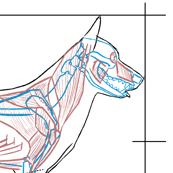
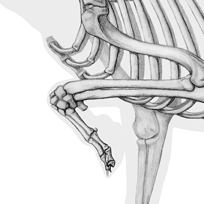
The musculoskeletal system plays a central role in many veterinary contexts. Orthopedic conditions, such as cruciate ligament injuries, require clear visualization of joints, ligaments, and biomechanical forces. Without accurate representation, these relationships can be difficult to interpret.
Internal systems are equally complex. Cardiovascular, respiratory, and digestive structures must be represented with precision, particularly in educational and pharmaceutical contexts where clarity is essential.
Neurological structures, including both central and peripheral systems, are also frequently visualized in both research and clinical communication.
By combining anatomical knowledge with visual expertise, I translate complex systems into structured, understandable imagery that supports real-world applications.
The process: from concept to validated final image
Each project follows a structured and transparent workflow.
The process begins with a detailed briefing, during which the objectives, audience, and level of anatomical detail are defined. This is followed by a research phase that ensures all visual decisions are grounded in validated sources.
The concept stage focuses on structure, proportion, and composition. Once approved, the illustration is developed into a detailed final image, with attention to texture, color, and clarity.
Review moments are integrated throughout the process, allowing for validation before final delivery.
Final files are delivered in high resolution and appropriate formats, with clear agreements regarding usage and licensing.
Project timelines typically range from two to eight weeks, depending on complexity.
A transparent workflow designed for accuracy, clarity, and efficient collaboration.

From detailed illustration to 3D-supported canine visualization
Canine anatomy can be visualized in different ways depending on the purpose of the project. Some subjects are best communicated through precise 2D illustration, especially when the goal is clarity, labeling, education, or publication. Other projects may benefit from 3D-supported visualization when spatial relationships, joint mechanics, skeletal structures, or surgical anatomy need to be explored more clearly.
For canine anatomy, this can be useful when explaining regions such as the skull, spine, limbs, joints, musculature, organ systems, or neurological structures. A 3D approach can support understanding, but it should always serve the communication goal rather than become a technical effect on its own.
Depending on the project, the visual approach may include detailed line illustration, digital painting, vector-based explanatory visuals, or selected 3D visualization. The choice of technique is guided by the audience, anatomical complexity, and final use.
Related services include:
Applications across veterinary, academic, and canine health communication
Canine anatomy illustration is used in veterinary education, clinical communication, research, publishing, and animal health communication. Because dogs show major variation in body type, skull shape, limb proportions, and breed-related anatomy, clear visual explanation can be especially valuable.
In veterinary education, canine visuals help students understand structures that are difficult to interpret from dissections, diagnostic images, or photographs alone. In clinical communication, illustrations can support explanation of orthopedic conditions, neurological pathways, surgical procedures, organ systems, or treatment plans.
For research and publishing, canine anatomy illustration can clarify anatomical relationships, breed-specific morphology, comparative studies, and medical or scientific findings. The goal is always to create visuals that are accurate, calm, and useful for professional communication.
Why custom illustration outperforms stock and AI-generated imagery
There are many visual solutions available today, including stock platforms, online tools, and AI-generated imagery. However, these options rarely meet the standards required for professional veterinary use.
Stock images are often generic and cannot be adapted to specific anatomical or clinical contexts. Template-based tools lack depth and precision. AI-generated images, while improving, still introduce significant risks, particularly in anatomy, where small errors can have major implications.
Custom canine anatomy illustration offers full control over accuracy, detail, and presentation. Each visual is tailored to the specific context, audience, and purpose.
In addition, licensing is clear and structured, ensuring the work can be used with confidence across professional applications.
For organizations working at a high level, reliability and accuracy are essential and cannot be outsourced to generic solutions.
About Karin Spijker
I am Karin Spijker, a scientific and medical illustrator with a background in drawing, painting, and textile design. With a master’s in scientific illustration and additional skills in 3D production, I combine accuracy, artistry, and storytelling. My work helps healthcare and publishing teams communicate complex ideas clearly and reliably.
Alongside commissioned projects, I also create independent artworks inspired by nature, anatomy, and landscapes. My mission is to make science and nature accessible, inspiring, and visually engaging.

Who canine anatomy illustration is for
Canine anatomy illustration is useful for professionals and organizations that need to communicate dog anatomy with accuracy, structure, and visual clarity. This may include veterinary educators, clinicians, researchers, publishers, animal health organizations, dog-related educational platforms, and communication teams working with veterinary or scientific content.
In education, canine anatomy visuals can help students understand structures that are difficult to interpret from photographs, dissections, or diagnostic images alone. In clinical and client communication, they can help explain conditions, procedures, or anatomical regions in a calm and understandable way.
The focus is not on creating decorative dog artwork, but on translating canine anatomy into visuals that are accurate, useful, and professionally prepared.
Related pages:
→ animal anatomy illustration
→ medical illustration services
→ patient education illustration


Understanding canine anatomy illustration pricing
Every project is custom and depends on scope, complexity, and intended usage. If you would like a clear overview of how pricing is structured, including typical ranges and licensing, you can explore the pricing page.
Frequently asked questions
Find answers to common questions about the illustration process, timelines, pricing, licensing, file delivery, and collaboration.
Work with Karin Spijker
A well-executed canine anatomy illustration strengthens communication, improves understanding, and enhances the credibility of your work.
Whether you are developing educational materials, conducting research, or working on clinical communication, high-quality visualization plays a key role.
A professional canine anatomy illustration transforms complex anatomical information into clear, structured, and reliable visual communication.
It supports better decision-making, stronger education, and more effective collaboration, making it an essential tool within modern veterinary and scientific practice.
You can:
→ view the portfolio
→ request a quote via medical illustration services
→ or get in touch to discuss your project
Translate complex canine anatomy into clear visual communication
Work directly with a scientific and medical illustrator to create accurate visuals of canine anatomy for veterinary education, research, publication, or clinical communication.
Ready to collaborate on a canine anatomy illustration project?
Clean, accurate, and aligned with your anatomical content.
