
Comparative anatomical accuracy
Illustrations developed from anatomical references, scientific source material, and species-specific structures.
Clarity across species
Visuals designed to compare anatomical similarities, differences, adaptations, and functional relationships.
Structured visual comparison
A clear workflow from research question and reference material to final visuals for publication, education, or veterinary use.
Comparative anatomy illustration services
Comparative anatomy illustration translates anatomical similarities and differences between species into clear, accurate visual communication. In evolutionary biology, zoology, veterinary science, medical research, and education, small structural differences can carry significant scientific meaning. A well-developed illustration helps make those differences visible, understandable, and useful.
Comparative anatomy often involves complex relationships between form, function, development, and evolutionary history. Text alone can describe these relationships, but it rarely conveys them with the immediacy and clarity that a structured visual can. Illustration allows multiple species, structures, or systems to be shown side by side with consistent perspective, hierarchy, labeling, and visual logic.
I am Karin Spijker, a certified scientific and medical illustrator with a strong background in anatomy, biological visualization, and detailed observational drawing. I create comparative anatomy illustrations for research, education, scientific publishing, veterinary communication, and life science projects.
Related pages:
→ anatomy illustration
→ animal anatomy illustration
→ scientific illustration
→ portfolio
“I have worked with Karin Spijker for many years on various visual projects. The greatest common denominator in these projects is a qualitative, professional image delivered on time. Karin is entirely at home in both fields, whether an illustration or high-end image editing on photo material.”
“Karin Spijker has performed assignments for me several times to my complete satisfaction, such as logos and two 3D animations. Karin can translate the information from a briefing into the desired end product and can think along with you pleasantly. In doing so, she works accurately, follows the set timetable, and honors her appointments. Karin is also a charming person to work with.”
 Natasja Kardos, Seahorse Solutions
“For my clients, I have asked Karin Spijker more often for customized assignments, especially for more specialized image editing. Karin can conjure up software like Adobe Photoshop and Illustrator. She is very meticulous and also communicates about progress. I highly recommend Karin, her work, and her pleasant cooperation.”
 Marian de Jong, Piri Piri Marketing en Communicatie
Turning comparative anatomy into visual clarity
Comparative anatomy is the study of anatomical relationships across species. It examines how structures are shared, adapted, modified, or specialized in different organisms. These comparisons can reveal evolutionary patterns, functional adaptations, developmental relationships, or clinically relevant similarities between animal and human anatomy.
Illustration is especially valuable in this field because it can organize complex biological information into a coherent visual system. Instead of presenting isolated images, a comparative anatomy illustration can align structures, standardize viewpoints, and clarify relationships that would otherwise remain difficult to compare.
For example, skeletal structures, muscle attachments, organ systems, vascular patterns, or limb adaptations can be visualized in a way that allows immediate comparison. The goal is not to make different species look artificially similar, but to clearly and accurately show meaningful anatomical relationships.
Why comparative anatomy illustration matters
In comparative anatomy, subtle differences matter. A small variation in bone shape, joint structure, muscle attachment, organ placement, or proportional relationship can influence how a species moves, feeds, breathes, reproduces, or adapts to its environment. These details are often difficult to communicate through photography, diagrams, or written description alone.
A well-designed comparative anatomy illustration supports scientific communication by showing what is similar, what is different, and why those differences matter. It can help researchers explain their findings, educators teach complex biological concepts, and publishers present anatomical information in a clear, visually consistent way.
This is particularly useful in contexts such as veterinary education, evolutionary biology, zoology, biomechanics, natural history, and translational research, where anatomical comparison is central to the message.
Related pages:
→ veterinary medical illustration
→ natural history illustration
→ biological illustration
Morphological variation and scientific precision
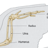
Comparative anatomy depends on precision. Homologous structures, analogous adaptations, and species-specific variations need to be represented carefully. A forelimb, skull, pelvis, fin, wing, or vertebral structure may appear visually similar across species, but its function and anatomical organization can differ significantly.
Scientific accuracy is therefore essential. Comparative anatomy illustrations may be developed from anatomical literature, specimen references, imaging data, dissection references, research papers, or expert input. The final visual must remain faithful to the source material while making the comparison easier to understand.
This balance is important. If too much detail is removed, the scientific meaning may be lost. If too much information is shown at once, the comparison becomes visually confusing. The role of the illustrator is to create a clear visual structure that supports interpretation without distorting the biology.
Workflow: from research question to final illustration
A comparative anatomy project begins with understanding the purpose of the comparison. Before drawing begins, the subject, audience, scientific message, level of detail, and final use must be clearly defined.
Reference material is then reviewed. This may include anatomical literature, published research, specimen photographs, CT or MRI data, sketches, diagrams, or input from subject-matter experts. The concept phase establishes the visual structure: which species or structures are shown, how they are aligned, what level of detail is needed, and how the viewer should read the comparison.
- briefing and scope definition
- reference review and anatomical research
- concept sketches and visual planning
- refinement based on feedback
- final illustration delivery for publication, education, or digital use
A transparent workflow designed for accuracy, clarity, and efficient collaboration.

From raw data to structured visual insight
A comparative anatomy illustration is not simply a set of animal drawings placed next to each other. It is a visual interpretation of biological information. Each project begins with a clear communication goal: what needs to be compared, why the comparison matters, and who needs to understand it.
Some projects focus on evolutionary relationships, such as skeletal adaptations across vertebrates. Others may compare functional anatomy, such as limb mechanics, jaw structures, respiratory systems, or cardiovascular organization. In veterinary or biomedical contexts, comparative anatomy may also help explain similarities and differences between animal models and human anatomy.
The illustration process brings these references together into a structured visual explanation. Viewpoints may be standardized, labels refined, unnecessary visual noise removed, and key structures emphasized. This creates a visual that balances scientific accuracy with readability.
Comparative anatomy for research and publication
In research and scientific publishing, comparative anatomy illustrations help transform complex data into visuals that can be interpreted quickly and accurately. They are useful when a study depends on structural comparison, species variation, morphology, functional anatomy, or evolutionary interpretation.
A clear comparative visual can strengthen a publication by making the research question easier to understand. It can also help reviewers, readers, and interdisciplinary audiences follow the work’s anatomical logic. This is especially valuable when the subject involves specialized terminology, complex spatial relationships, or multiple species.
- peer-reviewed publications
- scientific figures and diagrams
- textbooks and educational materials
- zoological and veterinary research
- evolutionary biology communication
- conference presentations and lectures
→
About Karin Spijker
I am Karin Spijker, a scientific and medical illustrator with a background in drawing, painting, and textile design. With a master’s in scientific illustration and additional skills in 3D production, I combine accuracy, artistry, and storytelling. My work helps healthcare and publishing teams communicate complex ideas clearly and reliably.
Alongside commissioned projects, I also create independent artworks inspired by nature, anatomy, and landscapes. My mission is to make science and nature accessible, inspiring, and visually engaging.

Comparative anatomy for education
Comparative anatomy is inherently visual. Students often need to understand how one structure relates to another across species, and why those similarities or differences matter. Illustrations can make this learning process more efficient by visually organizing the information.
A strong comparative anatomy illustration can clarify structural relationships that are difficult to understand from separate photographs or specimen images. It can show consistent orientation, proportional relationships, labeling systems, and visual hierarchy across multiple animals or anatomical systems.
This makes comparative anatomy illustration valuable for veterinary schools, biology programs, zoology courses, e-learning platforms, museum education, and scientific outreach. A well-made visual can be reused across lectures, handouts, textbooks, digital platforms, and long-term educational materials.
Practical applications of comparative anatomy illustration
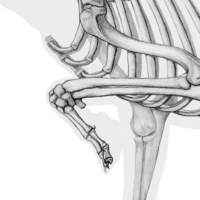
Comparative anatomy illustrations can be used across a wide range of scientific and educational contexts. For example, a visual comparison of vertebrate forelimbs may help explain how similar skeletal structures are adapted for walking, flying, swimming, or grasping. A comparison of skull morphology may reveal differences in feeding strategy, sensory specialization, or evolutionary adaptation.
In veterinary and biological education, comparative visuals can help clarify relationships between domestic animals, wildlife, and human anatomy. In research contexts, they can support the explanation of morphology, biomechanics, species adaptation, or anatomical variation.
- skeletal comparisons
- muscle and tendon relationships
- organ system variation
- evolutionary adaptations
- comparative veterinary anatomy
- functional anatomy and biomechanics
- human-animal anatomical comparison
Custom comparative anatomy illustration versus generic visuals
Stock images, template tools, and AI-generated visuals are rarely suitable for serious comparative anatomy work. They may be useful for rough inspiration or simple communication, but they often lack the anatomical precision and species-specific accuracy required for research, education, and publication.
Comparative anatomy requires control over viewpoint, scale, proportion, labeling, detail, and interpretation. Generic visuals usually cannot provide this level of consistency. Even when individual images are accurate, they may not be suitable for direct comparison because they were not created within the same visual system.
- species-specific anatomical accuracy
- consistent viewpoints and visual hierarchy
- control over detail, labeling, and emphasis
- alignment with your research or educational purpose
- publication-ready output and clear usage rights

Techniques and output formats
The technique depends on the project’s goal. Some comparative anatomy visuals require clean line work and clear labeling, while others benefit from detailed digital rendering, vector illustration, or 3D visualization. The visual approach is selected based on clarity, accuracy, audience, and final use.
For scientific publications, visuals can be prepared as high-resolution raster or vector files. For teaching and presentations, illustrations may be adapted for slides, handouts, posters, or digital platforms. For more complex structures, 3D visualization may support spatial understanding.
The final files can be prepared for journal submission, educational use, print production, presentations, or online communication.
Understanding comparative anatomy illustration pricing
Every project is custom and depends on scope, complexity, and intended usage. If you would like a clear overview of how pricing is structured, including typical ranges and licensing, you can explore the pricing page.
Why work with Karin Spijker
Comparative anatomy illustration requires more than the ability to draw animals. It requires an understanding of anatomy, scientific interpretation, visual discipline, and the ability to translate complex biological relationships into clear communication.
My work combines medical and scientific illustration training with strong observational drawing skills and a deep interest in anatomy, animals, and natural forms. This makes comparative anatomy a natural bridge between my medical, scientific, and nature-based work.
Projects are developed with attention to accuracy, clarity, collaboration, and usability. Whether the final illustration is intended for research, education, publication, or veterinary communication, the aim is to create visuals that support understanding and can be used with confidence.
Long-term value of comparative anatomy illustration
A well-developed comparative anatomy illustration is not just a one-time visual. It can become part of a larger scientific or educational communication system. The same visual approach can be adapted across publications, lectures, textbooks, digital learning environments, museum displays, and future project materials.
Consistency is especially important in comparative work. When multiple species or structures are visualized together, the style, perspective, labeling, and level of detail must remain consistent. This makes the final result more professional, easier to understand, and more useful over time.
Custom comparative anatomy illustration supports clarity, reliability, and long-term communication value.
Frequently asked questions
Find answers to common questions about the illustration process, timelines, pricing, licensing, file delivery, and collaboration.
Start your comparative anatomy illustration project
If you are looking for a comparative anatomy illustrator for research, education, publication, or veterinary communication, the focus should always be on clarity, accuracy, and usability.
Whether your project involves species comparison, evolutionary anatomy, veterinary education, functional morphology, or broader scientific communication, I can help translate complex anatomical relationships into clear, reliable, and professionally prepared visuals.
→ View related work in the portfolio
→ Get in touch to discuss your project
