
Orthopedic and anatomical accuracy
Illustrations developed from validated anatomical references, imaging, and procedural source material.
Clarity for complex musculoskeletal communication
Structured visuals designed to explain anatomy, procedures, biomechanics, and reconstruction clearly.
Publication-ready visualization
Medical visuals created for surgery, education, research, and clinical communication.
Orthopedic medical illustration
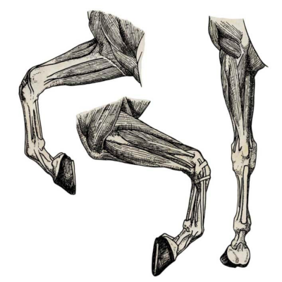
Orthopedic medical illustration translates complex musculoskeletal anatomy into clear, scientifically accurate visual communication for surgery, research, education, and clinical use. Orthopedics involves highly detailed anatomical relationships between bones, joints, ligaments, tendons, muscles, implants, and surrounding neurovascular structures. Communicating these accurately is essential in environments where precision directly affects interpretation, decision-making, and patient outcomes.
Standard photography, operative footage, or generic stock visuals often fail to provide the clarity required for orthopedic communication. Surgical procedures may involve overlapping structures, limited visibility, complex spatial relationships, or highly technical procedural steps that are difficult to interpret quickly from imaging alone. Medical illustration introduces hierarchy, structure, and selective emphasis to clarify what matters most.
I am Karin Spijker, a certified scientific and medical illustrator specializing in anatomy, biomedical visualization, and complex medical communication. I create custom orthopedic medical illustrations designed for surgical communication, scientific publishing, education, patient explanation, and publication-ready visualization.
Related pages:
→ medical illustration
→ surgical illustration
→ medical animation
“I have worked with Karin Spijker for many years on various visual projects. The greatest common denominator in these projects is a qualitative, professional image delivered on time. Karin is entirely at home in both fields, whether an illustration or high-end image editing on photo material.”
“Karin Spijker has performed assignments for me several times to my complete satisfaction, such as logos and two 3D animations. Karin can translate the information from a briefing into the desired end product and can think along with you pleasantly. In doing so, she works accurately, follows the set timetable, and honors her appointments. Karin is also a charming person to work with.”
 Natasja Kardos, Seahorse Solutions
“For my clients, I have asked Karin Spijker more often for customized assignments, especially for more specialized image editing. Karin can conjure up software like Adobe Photoshop and Illustrator. She is very meticulous and also communicates about progress. I highly recommend Karin, her work, and her pleasant cooperation.”
 Marian de Jong, Piri Piri Marketing en Communicatie
Translating complex orthopedic anatomy into visual clarity
Orthopedic communication often depends on extremely precise anatomical interpretation. Even small differences in joint alignment, implant positioning, fracture configuration, or biomechanical balance can significantly influence both surgical outcomes and medical decision-making. Communicating these relationships clearly is not always possible through imaging, operative photography, or verbal explanation alone.
Medical illustration helps organize complex musculoskeletal information into structured visual communication that is easier to interpret without losing scientific accuracy.
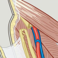
This is particularly valuable when visualizing fractures, osteotomies, tendon reconstruction procedures, implant positioning, degenerative joint conditions, and biomechanical relationships between bones, muscles, ligaments, and surrounding soft tissues. Many orthopedic procedures involve highly layered anatomy and complex spatial relationships that can become visually overwhelming in raw imaging or surgical footage.
Orthopedic medical illustration introduces hierarchy and selective emphasis by isolating the structures and procedural details that matter most for the intended audience. Rather than presenting every visible detail at once, the illustration focuses attention on anatomical relationships, surgical approaches, fixation systems, or movement mechanics that are essential for understanding the case.
This type of visualization supports communication across orthopedic surgery, education, scientific publishing, implant development, patient communication, and interdisciplinary collaboration. The goal is not to simplify orthopedic reality, but to translate complex musculoskeletal anatomy into visuals that are clearer, more focused, and easier to understand.
Surgical precision and anatomical accuracy
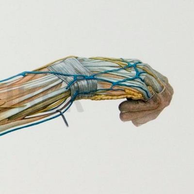
In orthopedic surgery, anatomical precision is directly connected to procedural understanding and surgical outcome. Procedures often involve highly complex spatial relationships between bones, joints, ligaments, tendons, muscles, implants, and surrounding neurovascular structures. Small inaccuracies in visualization or interpretation can significantly affect communication, planning, and education.
Medical illustration helps translate this complexity into structured visual communication that is clearer and easier to interpret than raw imaging or operative photography alone.
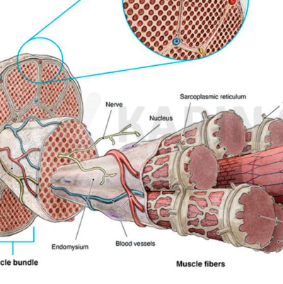
Orthopedic illustrations are frequently developed using medical source material such as MRI and CT imaging, X-rays, radiographic references, surgical documentation, and anatomical literature. From this information, anatomical structures can be reconstructed digitally layer by layer to accurately visualize fractures, osteotomies, implant positioning, fixation systems, tendon reconstruction, or biomechanical relationships.
This level of visualization is particularly valuable in orthopedic surgery and procedural planning, where illustrations may clarify surgical access routes, procedural workflows, implant orientation, or relationships among anatomical structures within confined operative fields.
In educational and scientific contexts, orthopedic medical illustration also supports clearer communication of complex procedures and musculoskeletal anatomy for residents, surgeons, researchers, publishers, and interdisciplinary teams.
The purpose of orthopedic illustration is not simply to depict anatomy in detail. Its role is to organize complex orthopedic information into visuals that are anatomically reliable, scientifically grounded, and easier to understand in both clinical and academic environments.
From medical data to final visualization
- review of imaging and medical source material
- alignment on goals, audience, and application
- concept sketches and anatomical planning
- scientific development and refinement
- final delivery in publication-ready formats
A transparent workflow designed for accuracy, clarity, and efficient collaboration.

Applications across orthopedic communication
Orthopedic medical illustration is used across a broad range of clinical, scientific, educational, and professional contexts where accurate visualization improves understanding and communication.
In surgical and clinical settings, illustrations help explain procedures, treatment strategies, anatomical relationships, and procedural workflows more efficiently than imaging alone. This can support preoperative planning, resident education, conference presentations, and interdisciplinary collaboration between healthcare professionals.
In research and publishing, orthopedic visuals help organize complex biomechanical or anatomical information into publication-ready figures that improve readability and clarity of communication. Detailed illustrations are frequently used in scientific journals, textbooks, educational platforms, and conference presentations.
- reconstructive orthopedic procedures
- sports injuries and ligament reconstruction
- spine and joint-related conditions
- trauma and fracture communication
- patient-focused educational materials
- orthopedic device and implant communication
About Karin Spijker
I am Karin Spijker, a scientific and medical illustrator with a background in drawing, painting, and textile design. With a master’s in scientific illustration and additional skills in 3D production, I combine accuracy, artistry, and storytelling. My work helps healthcare and publishing teams communicate complex ideas clearly and reliably.
Alongside commissioned projects, I also create independent artworks inspired by nature, anatomy, and landscapes. My mission is to make science and nature accessible, inspiring, and visually engaging.

Orthopedic illustration for research and education
- clarity of anatomical communication
- readability of scientific publications
- understanding of biomechanical relationships
- engagement in teaching and presentations
- interpretation of procedural workflows
- peer-reviewed publications
- surgical atlases and textbooks
- orthopedic training programs
- e-learning platforms and presentations
- biomechanical and implant research
Each visual is developed with attention to anatomical precision, educational clarity, and publication standards, ensuring long-term usability across multiple professional contexts.
Custom orthopedic illustration versus generic visuals
Generic stock visuals and simplified anatomical graphics often lack the specificity required for orthopedic communication. They may oversimplify anatomical relationships, contain inaccuracies, or fail to represent the exact procedure, implant, or biomechanical context involved.
- case-specific anatomical accuracy
- structured visual hierarchy
- procedural and biomechanical clarity
- publication-ready quality
- adaptability for revision and refinement
- consistent visual communication across materials
|
Feature
|
Custom Medical Illustrations (Karin Spijker)
|
Stock Images / AI Tools
|
|---|---|---|
|
Scientific accuracy
|
Based on validated medical references and anatomical expertise |
Often generic, outdated, or factually incorrect
|
|
Uniqueness
|
Built from validated medical source material |
Often generic, outdated, or factually incorrect
|
|
Didactic focus
|
Specifically designed to explain your message
|
General imagery, often lacking instructional focus
|
|
Flexibility
|
Fully adaptable throughout the design process
|
No or very limited customization
|
|
Visual consistency
|
A consistent visual language across materials
|
Mixed styles from multiple sources
|


Patient communication and medico-legal visualization
Orthopedic visuals are frequently used outside the operating room as well. In patient communication, clear illustrations help explain diagnoses, procedures, reconstruction techniques, or expected outcomes without relying solely on graphic operative photography or difficult-to-read imaging.
- informed consent discussions
- rehabilitation explanation
- treatment planning communication
- procedural understanding for patients and families
- evidence-based
- anatomically reliable
- visually clear
- professionally objective
→ medico-legal illustration
Understanding orthopedic medical illustration pricing
Every project is custom and depends on scope, complexity, and intended usage. If you would like a clear overview of how pricing is structured, including typical ranges and licensing, you can explore the pricing page.
Frequently asked questions
Find answers to common questions about the illustration process, timelines, pricing, licensing, file delivery, and collaboration.
Clear orthopedic communication through medical illustration
Orthopedic communication depends on clarity, structure, and anatomical precision. Whether the goal is surgical explanation, scientific publishing, education, patient communication, or medico-legal visualization, high-quality visuals improve understanding and support more effective communication.
