Nasal anatomical accuracy
Illustrations developed from verified medical references, surgical source material, and accurate nasal anatomy.
Clarity for rhinoplasty communication
Visuals designed to explain nasal structures, cartilage relationships, airway function, and surgical concepts clearly.
Structured visual communication
A clear workflow from briefing and reference material to final visuals for surgical education, publication, or patient communication.
Rhinoplasty medical illustration services
Rhinoplasty medical illustration translates complex nasal anatomy and surgical techniques into clear, accurate visual communication. Rhinoplasty is one of the most technically demanding procedures within aesthetic and reconstructive surgery. The nose is a small but highly complex three-dimensional structure, formed by bone, cartilage, mucosa, soft tissue, skin, and functional airway relationships.
Small anatomical changes can influence both external appearance and nasal function. This makes visual clarity essential, especially when a procedure needs to be explained to surgeons, students, publishers, patients, or professional audiences.
I am Karin Spijker, a certified scientific and medical illustrator with a strong background in anatomy, surgical visualization, and detailed drawing. I create custom rhinoplasty medical illustrations for surgical education, academic publications, clinical communication, patient education, and professional presentation material.
Related pages:
→ medical illustration
→ surgical illustration
→ plastic surgery medical illustration
→ reconstructive surgery illustration
→ patient education illustration
→ portfolio
“I have worked with Karin Spijker for many years on various visual projects. The greatest common denominator in these projects is a qualitative, professional image delivered on time. Karin is entirely at home in both fields, whether an illustration or high-end image editing on photo material.”
 Pauline Speelman, Signorita
“Karin Spijker has performed assignments for me several times to my complete satisfaction, such as logos and two 3D animations. Karin can translate the information from a briefing into the desired end product and can think along with you pleasantly. In doing so, she works accurately, follows the set timetable, and honors her appointments. Karin is also a charming person to work with.”
 Natasja Kardos, Seahorse Solutions
“For my clients, I have asked Karin Spijker more often for customized assignments, especially for more specialized image editing. Karin can conjure up software like Adobe Photoshop and Illustrator. She is very meticulous and also communicates about progress. I highly recommend Karin, her work, and her pleasant cooperation.”
 Marian de Jong, Piri Piri Marketing en Communicatie
Translating nasal anatomy into visual clarity
Rhinoplasty is difficult to communicate through photography alone. Intra-operative images can be useful as reference material, but they often contain blood, reflections, limited visibility, instruments, and visual distractions. They also cannot readily reveal the internal relationships among skin, cartilage, bone, mucosa, and airway structures.
Medical illustration makes it possible to clarify these relationships. It can show the nasal skeleton beneath the skin, isolate cartilage structures, explain the septum and nasal valves, or visualize the relationship between surgical modification and external form. By removing unnecessary visual noise and emphasizing what matters most, illustration helps the viewer understand the anatomy and the surgical logic behind the procedure.
The goal is not to simplify rhinoplasty into a generic diagram. The goal is to make complex nasal anatomy and surgical communication more understandable without losing anatomical accuracy.
Why rhinoplasty illustration matters
Rhinoplasty sits at the intersection of anatomy, aesthetics, function, and surgical decision-making. A visual explanation must therefore be precise and carefully structured. It is not enough to show the outside of the nose. The viewer often needs to understand what happens beneath the skin and how internal anatomical changes affect both shape and breathing.
A well-developed rhinoplasty illustration can clarify relationships that are difficult to explain in words. It may show the nasal bones, upper lateral cartilages, lower lateral cartilages, septum, nasal valves, dorsum, tip support mechanisms, or surgical planes in a readable, calm way.
This is valuable for surgical education, peer-reviewed publications, patient communication, conference presentations, books, websites, and professional teaching materials. In all of these contexts, the visual should support understanding rather than overwhelm the viewer.
Nasal anatomy, structure, and surgical communication
Nasal anatomy is highly layered. The external shape of the nose is influenced by internal support structures, cartilage frameworks, bony anatomy, skin thickness, soft tissue, mucosa, and airway function. These relationships are difficult to capture in a single photograph.
Illustration can show the nose layer by layer. It can separate external appearance from internal anatomy, clarify how cartilage structures support the nasal tip, show the relationship between the dorsum and septum, or explain how nasal valve function relates to airway patency.
For professional communication, this is especially useful when the subject involves structural rhinoplasty, preservation approaches, graft placement, osteotomies, septal correction, tip refinement, or functional airway considerations. The level of detail can be adapted to the audience, from highly technical surgical visuals to more accessible patient education illustrations.
Related pages:
→ human anatomy illustration
→ surgical illustration
→ medical illustration
From reference material to final illustration
A rhinoplasty medical illustration project begins with the communication goal. Before drawing starts, it is important to define what needs to be shown, who the audience is, and where the final visual will be used.
Reference material may include surgical sketches, operative photographs, anatomical literature, imaging, existing diagrams, publication drafts, or expert input. The concept phase establishes composition, viewpoint, level of detail, and visual emphasis. Once the structure is approved, the final illustration is developed with attention to anatomy, clarity, color, labeling, and format.
- briefing and scope definition
- review of reference material
- concept sketches and visual planning
- illustration development and refinement
- final delivery for publication, presentation, web, or patient education use
A transparent workflow designed for accuracy, clarity, and efficient collaboration.

Rhinoplasty illustration for surgical education and publications
Surgical education requires clarity, sequence, and anatomical precision. A rhinoplasty technique is often understood not from a single image but from a structured visual explanation. Medical illustration can show what is exposed, what is preserved, what is modified, and how each step relates to the final surgical goal.
For textbooks, surgical atlases, journal figures, conference presentations, and teaching materials, rhinoplasty illustrations can support explanations of procedures such as dorsal modification, cartilage reshaping, septal support, graft placement, osteotomy lines, nasal valve correction, or tip suturing. The illustration can focus attention on the relevant structures while reducing distraction from surrounding tissue.
A strong surgical illustration does more than show “what happened.” It helps explain why the technique works and how the anatomical structures relate to one another.
Related pages:
→ surgical illustration
→ scientific illustration
Rhinoplasty illustration for patient education
Rhinoplasty patients often need a clear explanation, but overly graphic surgical photography can be confronting or confusing. Illustrations offer a calmer and more controlled way to explain anatomy, procedures, and expected changes.
Patient education visuals can help explain the difference between external appearance and internal structure. They can show how cartilage, bone, septum, or airway structures are involved in a procedure without becoming too technical or visually overwhelming.
This is useful for consultation materials, clinic websites, patient brochures, informed consent support, and educational communication. The visual language can be simplified without becoming inaccurate. The aim is to help patients understand the procedure more clearly and feel better informed.
Related page:
→ patient education illustration
Aesthetic and reconstructive rhinoplasty communication
Rhinoplasty may be performed for aesthetic, reconstructive, or functional reasons, and often these concerns overlap. Medical illustration can help communicate this complexity by showing the anatomical foundation behind visible form.
In aesthetic rhinoplasty, illustration may clarify dorsal shape, tip support, cartilage relationships, profile changes, or proportional balance. In reconstructive or functional contexts, it may help explain structural support, airway function, trauma-related changes, or the relationship between anatomy and breathing.
The visual approach should match the purpose of the communication. A publication figure may require precise anatomical labeling. A clinic website may need a refined but accessible explanatory visual. A patient education image may focus on calm clarity. In each case, accuracy and readability remain the foundation.
Related pages:
→ plastic surgery medical illustration
→ reconstructive surgery illustration
→ medical artwork
About Karin Spijker
I am Karin Spijker, a scientific and medical illustrator with a background in drawing, painting, and textile design. With a master’s in scientific illustration and additional skills in 3D production, I combine accuracy, artistry, and storytelling. My work helps healthcare and publishing teams communicate complex ideas clearly and reliably.
Alongside commissioned projects, I also create independent artworks inspired by nature, anatomy, and landscapes. My mission is to make science and nature accessible, inspiring, and visually engaging.

Custom rhinoplasty medical illustration versus generic visuals
- anatomically accurate nasal visualization
- clear surgical or educational focus
- controlled simplification without distortion
- publication-ready quality
- consistent style across larger projects
Techniques and output formats
The technique depends on the project’s goal. Some rhinoplasty illustrations are best communicated through clean line work or schematic diagrams. Others require detailed digital rendering, layered anatomical views, cross-sections, or selected 3D-supported visualization when spatial relationships need to be explained more clearly.
For scientific publishing, illustrations can be prepared as high-resolution raster or vector artwork. For presentations and patient education, visuals may be adapted for slides, clinic websites, brochures, digital learning materials, or professional communication. When needed, layered files can also support future revisions or related educational adaptations.
The choice of technique is guided by accuracy, audience, final use, and the level of anatomical detail required.


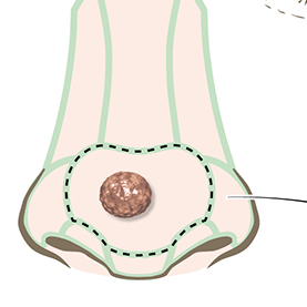

Understanding rhinoplasty medical illustration pricing
Rhinoplasty medical illustration is custom work. Pricing depends on the complexity of the subject, the number of views, the level of anatomical detail, the intended use, and the licensing scope.
A single explanatory illustration is different from a multi-step surgical sequence or a detailed anatomical plate. A patient education visual may require a different level of detail than a journal figure or textbook illustration. Licensing also matters: visuals used in a single publication have a different scope from visuals used across websites, lectures, printed material, marketing, and long-term educational resources.
After a short intake, I provide a clear quotation with scope, deliverables, timeline, usage rights, and revision structure. This ensures that expectations are clear before production begins.
Why work with Karin Spijker
Rhinoplasty medical illustration requires more than general drawing ability. It requires anatomical understanding, visual discipline, surgical awareness, and the ability to translate complex three-dimensional structures into clear visual communication.
My work combines medical and scientific illustration training with detailed anatomical drawing and a calm, structured visual language. I focus on visuals that are accurate, readable, and professionally prepared for their intended use.
Projects are developed with attention to clarity, collaboration, and usability. Whether the final illustration is intended for surgical education, academic publication, patient communication, or professional presentation, the aim is to create visuals that support understanding and can be used with confidence.
Long-term value of a custom rhinoplasty illustration
A strong rhinoplasty illustration can be used beyond a single project. The same visual approach may support journal figures, lectures, clinic education, website content, patient materials, or future teaching resources.
Consistency is especially valuable when multiple illustrations are created for the same publication, clinic, procedure, or educational system. A coherent visual language improves readability, supports learning, and gives the project a more professional appearance.
Custom rhinoplasty medical illustration supports clarity, accuracy, credibility, and long-term communication value.
Frequently asked questions
Find answers to common questions about the illustration process, timelines, pricing, licensing, file delivery, and collaboration.
Start your rhinoplasty medical illustration project
If you are looking for rhinoplasty medical illustration for surgical education, publication, patient communication, clinic materials, or professional presentations, the focus should always be on clarity, accuracy, and usability.
Whether your project involves nasal anatomy, cartilage structure, surgical technique, airway function, patient education, or a broader plastic surgery communication project, I can help translate complex rhinoplasty information into visuals that are clear, reliable, and professionally prepared.
→ View related work in the portfolio
→ Get in touch to discuss your project
Translate complex nasal anatomy into clear visual communication
Work directly with a scientific and medical illustrator to create accurate rhinoplasty visuals for surgical education, publication, patient communication, or professional presentations.
Ready to collaborate on a rhinoplasty medical illustration project?
Clear, accurate, and aligned with your medical content