
Equine anatomical accuracy
Illustrations developed from veterinary references, anatomical source material, and species-specific equine structures.
Clarity for complex equine anatomy
Visuals designed to explain musculoskeletal systems, biomechanics, movement, and clinical relationships clearly.
Structured visual communication
A clear workflow from briefing and reference material to final visuals for veterinary education, research, or publication.
Equine anatomy illustration services
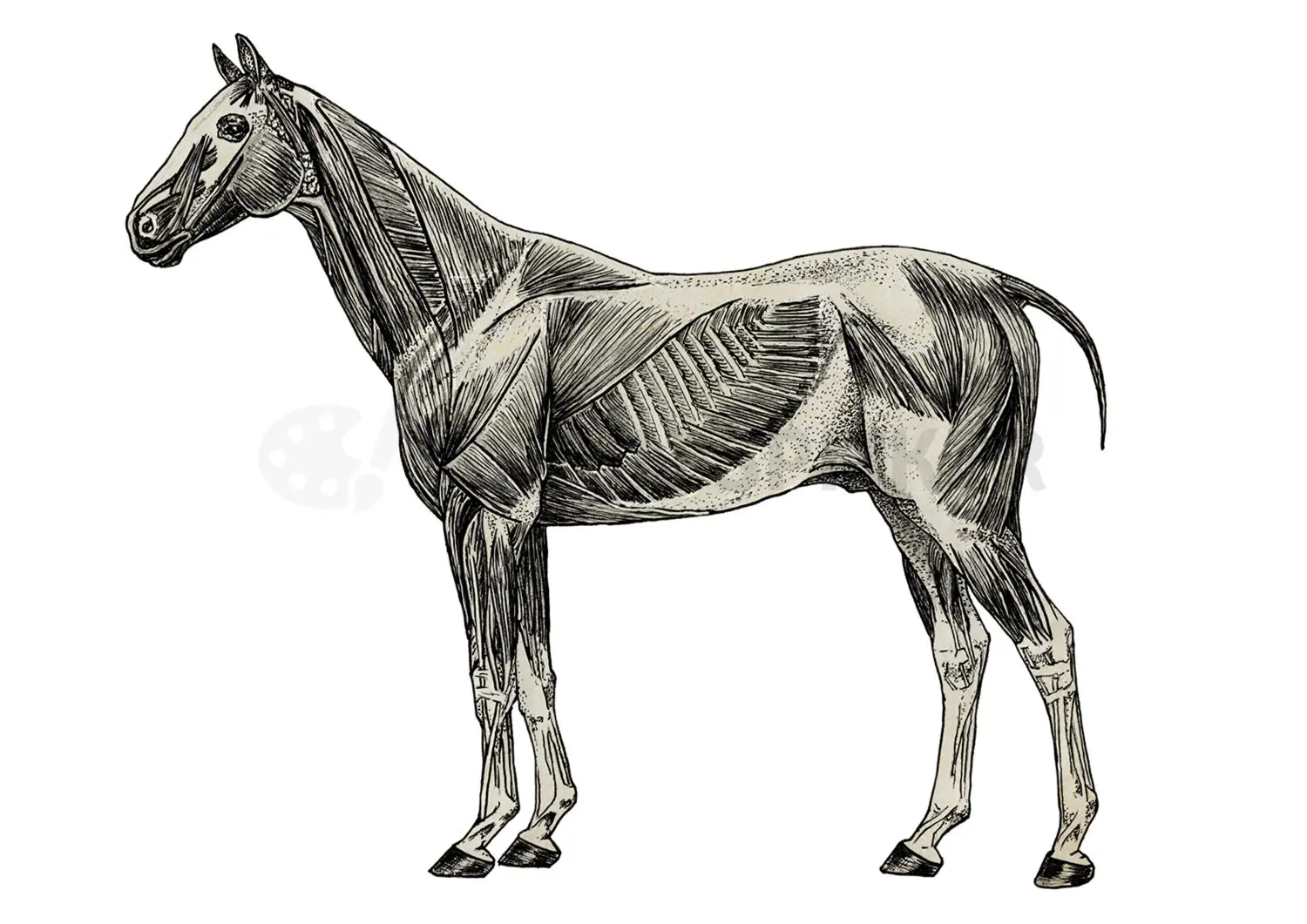
Equine anatomy illustration translates complex anatomical information about the horse into clear, accurate visual communication for veterinary education, research, clinical explanation, and publication. The equine body is highly specialized, with anatomical relationships that are closely connected to movement, performance, posture, and function.
A well-developed equine anatomy illustration can clarify structures that are difficult to explain through photography, diagnostic imaging, or text alone. This may include the musculoskeletal system, tendons and ligaments, hoof anatomy, joint structures, vascular relationships, organ systems, or anatomical regions involved in clinical or educational communication.
I am Karin Spijker, a certified scientific and medical illustrator with a strong background in anatomy, drawing, and biological visualization. I create custom equine anatomy illustrations for veterinary communication, educational materials, scientific publications, and research-based visual projects.
Related pages:
→ animal anatomy illustration
→ veterinary medical illustration
→ comparative anatomy illustration
→ portfolio
“I have worked with Karin Spijker for many years on various visual projects. The greatest common denominator in these projects is a qualitative, professional image delivered on time. Karin is entirely at home in both fields, whether an illustration or high-end image editing on photo material.”
“Karin Spijker has performed assignments for me several times to my complete satisfaction, such as logos and two 3D animations. Karin can translate the information from a briefing into the desired end product and can think along with you pleasantly. In doing so, she works accurately, follows the set timetable, and honors her appointments. Karin is also a charming person to work with.”
 Natasja Kardos, Seahorse Solutions
“For my clients, I have asked Karin Spijker more often for customized assignments, especially for more specialized image editing. Karin can conjure up software like Adobe Photoshop and Illustrator. She is very meticulous and also communicates about progress. I highly recommend Karin, her work, and her pleasant cooperation.”
 Marian de Jong, Piri Piri Marketing en Communicatie
Why equine anatomy illustration is critical in veterinary practice and research
The anatomy of the horse is uniquely adapted for strength, endurance, and locomotion. From the musculoskeletal system to cardiovascular efficiency, every structure is optimized for performance under high physical demand.
This level of complexity makes equine anatomy particularly challenging to communicate. Text-based explanations alone are often insufficient to fully convey spatial relationships, biomechanical forces, and functional interactions.
A well-developed equine anatomy illustration provides the clarity that written information cannot. It translates complex anatomical structures into accessible visual insight, enabling faster understanding and more precise communication.
In equine veterinary medicine, where diagnostics, treatment, and performance analysis often intersect, this level of visual accuracy is essential.
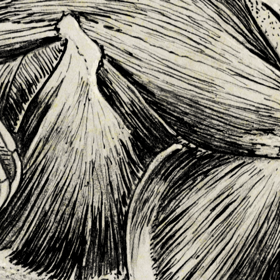
Scientific accuracy as the foundation of equine illustration
- anatomical proportions at scale
- spatial relationships between structures
- correct veterinary terminology
- alignment with validated scientific references
In equine medicine, precision is especially critical. Even minor inaccuracies in joint structure, tendon positioning, or vascular pathways can lead to misunderstandings in education or clinical interpretation.
For this reason, every equine anatomy illustration is developed through a research-driven process. I work with anatomical atlases, scientific literature, and, where relevant, imaging data such as CT and MRI scans. Collaboration with equine specialists ensures that each visual meets the professional-use standards.
Understanding equine anatomy: scale, biomechanics, and function
The horse presents unique anatomical challenges due to its size, biomechanics, and functional demands.
The musculoskeletal system is central to equine health and performance. Structures such as tendons, ligaments, and joints must withstand significant forces during movement. Visualizing these relationships accurately is essential, particularly in orthopedic contexts.
Conditions such as tendon injuries, joint degeneration, or laminitis require precise anatomical representation to support both diagnosis and education.
The cardiovascular and respiratory systems are also highly specialized, supporting sustained physical exertion. Clear visualization of these systems is important in both research and pharmaceutical applications.
Additionally, the hoof structure, complex and biomechanically critical, is a frequent subject of equine anatomical illustration. Misrepresentation in this area is common in generic imagery and can lead to incorrect assumptions.
By combining anatomical expertise with visual clarity, I translate these complex systems into structured, understandable, and application-ready imagery.
The process: from briefing to validated final result
Each project follows a structured and transparent workflow.
It begins with a detailed briefing to define the objectives, audience, and level of anatomical detail. This is followed by a research phase that ensures all visual decisions are based on reliable sources.
The concept stage focuses on structure and proportion. Once approved, the illustration is developed into a refined final image, with attention to detail, texture, and clarity.
Review stages are integrated throughout the process, allowing for validation before final delivery.
Final files are delivered in high-resolution and appropriate formats, with clear licensing agreements.
Most projects are completed within two to eight weeks, depending on complexity.
A transparent workflow designed for accuracy, clarity, and efficient collaboration.

From detailed illustration to 3D-supported visualization
Equine anatomy can be visualized in different ways depending on the purpose of the project. Some subjects are best communicated through precise 2D illustration, especially when the goal is clarity, labeling, education, or publication. Other projects may benefit from 3D-supported visualization when spatial relationships, complex structures, or repeated viewpoints need to be explored more clearly.
For equine anatomy, this can be useful when explaining regions such as the hoof, joints, limbs, musculature, or internal anatomical systems where depth and orientation are important. A 3D approach can support understanding, but it should always serve the communication goal rather than become a technical effect in its own right.
Depending on the project, the visual approach may include detailed line illustration, digital painting, vector-based explanatory visuals, or selected 3D visualization. The choice of technique is always guided by the audience, the anatomical complexity, and the illustration’s final use.
Related pages:
→ biomedical illustration
→ scientific illustration
→ working process
Applications across the veterinary, academic, and equine industries
A professional equine anatomy illustration is used in a wide range of contexts.
In veterinary education, detailed visuals support the understanding of complex structures and biomechanics. Students benefit from clear representations of anatomical relationships that are difficult to grasp through text alone.
In research and scientific publishing, high-quality illustrations enhance clarity and support the communication of findings. Journals require visuals that meet strict scientific and formatting standards.
In clinical environments, illustrations improve communication between veterinarians, specialists, and clients. Complex diagnoses or treatment plans can be explained more effectively with accurate visual support.
In equine sports medicine and performance analysis, anatomical visualization is used to better understand movement, injury mechanisms, and recovery processes.
Pharmaceutical and biotech companies use equine anatomical visuals to explain mechanisms of action and connect internal processes to overall physiology.
Why custom equine illustration outperforms stock and AI
Although stock imagery, online tools, and AI-generated visuals are widely available, they rarely meet the requirements of professional equine use.
Stock images are typically generic and lack the specificity needed for equine anatomy. Online tools are limited in depth and flexibility. AI-generated visuals often contain anatomical inaccuracies, particularly in complex structures such as joints, tendons, and hooves.
A custom equine anatomy illustration offers full control over accuracy, detail, and communication. Each visual is tailored to the specific context, ensuring it effectively supports the intended message.
In addition, the licensing is clear and well-suited to professional use, allowing organizations to use the material with confidence.
For high-level veterinary, academic, and commercial applications, custom illustration remains the most reliable solution.
About Karin Spijker
I am Karin Spijker, a scientific and medical illustrator with a background in drawing, painting, and textile design. With a master’s in scientific illustration and additional skills in 3D production, I combine accuracy, artistry, and storytelling. My work helps healthcare and publishing teams communicate complex ideas clearly and reliably.
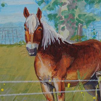
Alongside commissioned projects, I also create independent artworks inspired by nature, anatomy, and landscapes. My mission is to make science and nature accessible, inspiring, and visually engaging.

Who equine anatomy illustration is for
An equine anatomy illustration is useful for professionals and organizations that need to communicate horse anatomy accurately, with structure and visual clarity. This may include veterinary educators, equine specialists, researchers, publishers, animal health organizations, and communication teams working with veterinary or scientific content.
In education, equine anatomy visuals can help students understand structures that are difficult to interpret from photographs, dissections, or diagnostic images alone. In research and publishing, they can support a clear explanation of anatomical relationships, functional systems, or species-specific structures. In clinical and client communication, they can help explain conditions, procedures, or anatomical regions in a calm and understandable way.
The focus is not on creating decorative horse artwork, but on translating equine anatomy into visuals that are accurate, useful, and professionally prepared.
Related pages:
→ animal anatomy illustration
→ veterinary medical illustration
→ equine anatomy illustration
→ scientific illustration


Understanding equine anatomy illustration pricing
Every project is custom and depends on scope, complexity, and intended usage. If you would like a clear overview of how pricing is structured, including typical ranges and licensing, you can explore the pricing page.
Frequently asked questions
Find answers to common questions about the illustration process, timelines, pricing, licensing, file delivery, and collaboration.
Work with Karin Spijker
A well-executed equine anatomy illustration enhances clarity, supports understanding, and strengthens professional communication.
Whether you are working on education, research, or clinical applications, accurate visualization is a key component of effective communication.
A professional equine anatomy illustration transforms complex anatomical structures into clear, structured, and reliable visual communication.
It supports better learning, more precise clinical understanding, and stronger communication, making it an essential tool within modern equine veterinary and scientific practice.
- explore the portfolio
- request a quote via medical illustration services
- or get in touch to discuss your project
Translate complex equine anatomy into clear visual communication
Work directly with a scientific and medical illustrator to create accurate equine anatomy visuals for veterinary education, research, publication, or clinical communication.
Ready to collaborate on an equine anatomy illustration project?
Clean, accurate, and aligned with your anatomical content.
