
Neuroanatomical accuracy
Illustrations developed from validated references, imaging data, and evidence-based anatomical structures.
Clarity for complex neurological concepts
Structured visuals designed to simplify layered neuroanatomy and neurological communication.
Publication-ready visualization
Precise medical visuals for research, education, clinical communication, and scientific publishing.
Neurology medical illustration
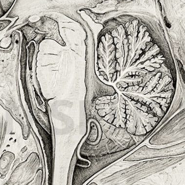
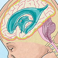
Neurology medical illustration translates complex neuroanatomy, neurological disorders, and neurosurgical concepts into clear visual communication for research, education, and clinical practice. The nervous system is one of the most complex systems in the human body, where small anatomical relationships can have major functional and clinical consequences. In these contexts, visual clarity is not optional. It is essential.
MRI scans, CT imaging, tractography, and surgical data often contain enormous amounts of information, but raw imaging alone rarely communicates efficiently to broader audiences. Medical illustration provides structure, hierarchy, and visual focus. Rather than showing everything at once, it isolates the information that matters most and translates it into understandable, publication-ready visuals.
I am Karin Spijker, a certified scientific and medical illustrator specializing in anatomy, biomedical visualization, and complex medical communication. I create custom neurology medical illustrations designed for scientific publishing, neurosurgical communication, medical education, and advanced healthcare visualization.
Related pages:
→ medical illustration
→ surgical illustration
→ medical animation
→ portfolio
“I have worked with Karin Spijker for many years on various visual projects. The greatest common denominator in these projects is a qualitative, professional image delivered on time. Karin is entirely at home in both fields, whether an illustration or high-end image editing on photo material.”
“Karin Spijker has performed assignments for me several times to my complete satisfaction, such as logos and two 3D animations. Karin can translate the information from a briefing into the desired end product and can think along with you pleasantly. In doing so, she works accurately, follows the set timetable, and honors her appointments. Karin is also a charming person to work with.”
 Natasja Kardos, Seahorse Solutions
“For my clients, I have asked Karin Spijker more often for customized assignments, especially for more specialized image editing. Karin can conjure up software like Adobe Photoshop and Illustrator. She is very meticulous and also communicates about progress. I highly recommend Karin, her work, and her pleasant cooperation.”
 Marian de Jong, Piri Piri Marketing en Communicatie
Translating neuroanatomy into visual clarity
- brain anatomy and neurovascular structures
- central and peripheral nervous system disorders
- neural pathways and tract systems
- neurosurgical procedures and approaches
- spinal anatomy and compression syndromes
- neurological mechanisms and treatment effects
Why visualization matters in neurology
- neurosurgical planning and communication
- university-level medical education
- scientific publishing and presentations
- patient communication and informed consent
- pharmaceutical and neurological pathway visualization
Neurology medical illustration for research and publication
- research publications and journal figures
- educational atlases and textbooks
- congress and conference presentations
- neurovascular and pathway visualization
- neurosurgical explanation and procedural support
- scientific communication and grant presentations
From medical concept to final illustration
- project briefing and scope definition
- review of medical and imaging source material
- concept sketches and visual planning
- refinement and feedback integration
- final production and delivery
A transparent workflow designed for accuracy, clarity, and efficient collaboration.

Clinical communication and patient understanding
- patient consultations
- informed consent discussions
- explanation of surgical procedures
- interdisciplinary communication
- educational handouts and presentations
In clinical settings, this improves understanding while reducing confusion and unnecessary anxiety.
Scientific accuracy and anatomical reliability
- validated anatomical references and atlases
- MRI, CT, and imaging-based source material
- scientific literature and clinical references
- subject-matter consultation when required
About Karin Spijker
I am Karin Spijker, a scientific and medical illustrator with a background in drawing, painting, and textile design. With a master’s in scientific illustration and additional skills in 3D production, I combine accuracy, artistry, and storytelling. My work helps healthcare and publishing teams communicate complex ideas clearly and reliably.
Alongside commissioned projects, I also create independent artworks inspired by nature, anatomy, and landscapes. My mission is to make science and nature accessible, inspiring, and visually engaging.

Techniques and visualization capabilities
- detailed 2D medical illustration
- 3D neuroanatomical modeling
- procedural and surgical visualization
- medical animation for neurological processes
- DICOM and imaging-based visualization
- compositing and visual refinement for publication
Each project is tailored to its final application, whether for publication, education, presentation, digital communication, or clinical use.
Custom neurology illustration versus generic visuals
- case-specific anatomical accuracy
- structured visual hierarchy
- alignment with research or clinical goals
- publication-ready quality
- flexibility for revision and refinement
|
Feature
|
Custom Medical Illustrations (Karin Spijker)
|
Stock Images / AI Tools
|
|---|---|---|
|
Scientific accuracy
|
Based on validated anatomical and scientific references |
Often generic, outdated, or factually incorrect
|
| Didactic focus | Specifically designed to explain your message | General imagery, often lacking instructional focus |
| Flexibility | Fully adaptable throughout the design process | No or very limited customization |


Understanding neurology medical illustration pricing
Every project is custom and depends on scope, complexity, and intended usage. If you would like a clear overview of how pricing is structured, including typical ranges and licensing, you can explore the pricing page.
Clear neurological communication through medical illustration
The quality of medical visualization directly influences how neurological information is understood. Whether the goal is scientific publishing, surgical communication, medical education, or patient explanation, clear visuals improve comprehension and support more effective communication.
Frequently asked questions
Find answers to common questions about the illustration process, timelines, pricing, licensing, file delivery, and collaboration.
